CILAB ← Back to projects

Project

Fast & Sustainable Brain MRI

Triple-modality deblurring for accelerated brain MRI — leveraging complementary information from orthogonally undersampled contrasts to shorten scan times and cut energy use, while preserving diagnostic image quality.

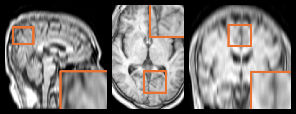
Overview of the triple-modality deblurring framework: T1, T2 and FLAIR are undersampled along mutually orthogonal phase-encoding directions, jointly registered, and each reconstructed by a deblurring network into a high-resolution image.
Overview of the proposed framework. Orthogonal, direction-specific undersampling is applied to each contrast (T1 in-plane horizontal, T2 in-plane vertical, FLAIR out-of-plane); after multi-contrast registration, the images are jointly reconstructed by a shared deblurring network. Source: project presentation (ISMRM 2025).
~4×
Acquisition acceleration over compressed sensing alone
60–75%
Reduction in gradient & RF energy per scan
0.84
Average SSIM across modalities
3
Contrasts accelerated jointly (T1, T2, FLAIR)

The MRI sustainability problem

Lengthy MRI acquisition negatively impacts sustainability through high energy consumption, increased patient discomfort, and reduced clinical efficiency. MRI is the single largest energy consumer in radiology, and its environmental footprint is growing on both fronts — more scans and more installed units.

Reducing acquisition duration directly addresses this: shorter sequences mean fewer gradient pulses, fewer RF cycles, and less cooling overhead — decreasing per-patient energy consumption, minimizing environmental impact, and improving scanner throughput.

Bar chart of approximate annual energy consumption per imaging system: MRI systems dominate at 100 MWh, versus 15 for C-arm, 10 for CT, 2 for X-ray and 1 for ultrasound.
MRI systems dwarf other imaging modalities in annual energy use. Average daily demand per unit fell from 226 kWh (2011) to 165 kWh (2017), yet total MRI energy still rose ≈15% as scan volumes and installed units grew. Data: Siemens Healthineers, 2023.

The limits of single-contrast reconstruction

Traditional acceleration techniques such as parallel imaging and compressed sensing successfully reduce acquisition times, but introduce reconstruction artifacts, offer limited gains at higher accelerations, and add computational demands. Recovering a scan from a single aggressively undersampled contrast is inherently ill-posed — auxiliary information is essential.

Prior multi-modal methods typically reconstruct accelerated contrasts using a reference contrast acquired at high resolution. We remove that dependency: we exploit complementary information from triplets of scans with mutually orthogonal phase-encoding directions, with no high-resolution priors and no vendor dependency.

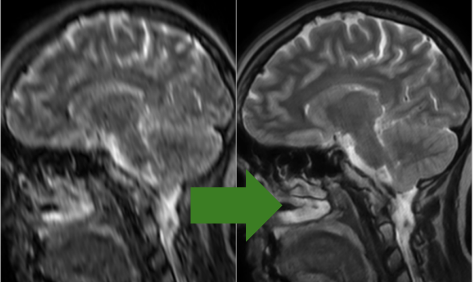
Pipeline showing a subsampled k-space producing a blurry scan that a deep-learning model reconstructs; the reconstruction from a single contrast remains imperfect.
Reconstructing from a single contrast is unreliable — the blur left by aggressive undersampling cannot be fully resolved without complementary information from other contrasts.

Our approach: orthogonal triple-modality undersampling

Acceleration is achieved by selectively reducing high-frequency components — undersampling roughly 3/4 of k-space along a distinct phase-encoding direction for each of the three contrasts (T1, T2, FLAIR). This shortens phase-encoding and readout durations, preserves essential low-frequency image contrast, reduces susceptibility artifacts, and substantially cuts gradient and RF energy consumption.

Because the blur direction differs per contrast, the missing high-frequency detail in one modality is captured by another. To exploit this, we first register the different contrasts to one another and then to an MNI atlas — improved spatial alignment significantly enhances network performance. Reconstruction then uses a custom deblurring network trained with a combined SSIM + L1 perceptual loss derived from VGG-19 features.

Orthogonal phase encoding turns three aggressively accelerated scans into a single complementary dataset — no contrast needs to be acquired at full resolution.

The reconstruction engine: FARD

Reconstruction is carried out by FARD — Frequency Attention Residual Denoising, a network designed for multi-contrast MRI deblurring with anisotropic kernels elongated in the direction of blur. FARD combines processing in both the frequency and image domains (global + local), and enforces sparsity in the attention domain — inspired by compressed sensing — so that attention favors anatomical structure over noise.

FARD architecture: main and auxiliary Conv2d branches are fused, passed through three FARD blocks and an output Conv2d with a residual connection; each block interleaves Conv, Attention and Frequency modules with decreasing then increasing channel counts.
FARD architecture. A main and an auxiliary convolutional branch are fused and refined through three FARD blocks, each interleaving convolution, attention, and frequency modules in a channel-narrowing then channel-widening path, with a global residual connection.

Results

The method was validated on healthy subjects scanned at 1.5T (Philips Ingenia), each undergoing both the accelerated protocol and clinical high-resolution scans as reference, and further validated on a large synthetically subsampled dataset (BRATS). Visual inspection consistently shows restoration of fine detail despite aggressive acceleration — and image quality improves monotonically as more complementary contrasts are added.

Grid of sagittal brain MRI reconstructions for T1 and T2 rows, comparing the accelerated single-contrast input against reconstructions using two and three modalities and the high-quality ground truth; magnified insets show progressively sharper detail.
Reconstruction quality grows with the number of complementary contrasts. Left to right: single-contrast input, two-modality, three-modality reconstruction, and high-quality ground truth. Magnified insets show recovery of fine anatomical structure as auxiliary contrasts are added.

Quantitatively, adding contrasts consistently raises both SSIM and PSNR for every target modality:

TargetModalities usedSSIM ↑ (mean/std)PSNR ↑ (mean/std)
T1T10.705 / 0.03324.71 / 0.74
T1 + T2 / T1 + FLAIR0.726 / 0.02225.05 / 0.66
T1 + T2 + FLAIR0.768 / 0.00226.31 / 0.00
T2T20.575 / 0.02222.19 / 0.58
T2 + T1 / T2 + FLAIR0.688 / 0.01625.51 / 0.40
T2 + FLAIR + T10.717 / 0.01025.95 / 0.57
FLAIRFLAIR0.614 / 0.01422.67 / 0.24
FLAIR + T2 / FLAIR + T10.644 / 0.01723.57 / 0.42
FLAIR + T1 + T20.696 / 0.01724.87 / 0.18
Cross-validated SSIM and PSNR by target contrast and set of modalities used. The full triple-contrast configuration (highlighted) is best for every target.

Acceleration reduces power consumption

The acceleration translates directly into sustainability gains on the scanner. For the full T1 + T2 + FLAIR protocol on a Philips 1.5T Ingenia, total acquisition drops from 10.4 minutes to 2.61 minutes, and total energy from 2.93 kWh to 0.72 kWh — up to a 75% reduction in energy per sequence.

Table of TE, TR, duration and energy for T1, T2 and FLAIR at regular versus fast settings on a Philips 1.5T Ingenia, showing large reductions in duration and energy for the accelerated scans.
Per-sequence acquisition time and energy, regular vs. accelerated, on a Philips 1.5T Ingenia. Duration and energy both fall sharply for every contrast, since the reconstruction works entirely in the image domain, post-acquisition.
Because reconstruction happens post-acquisition in the image domain, the method is scanner-agnostic — the energy and time savings come for free on existing hardware.

Summary & outlook

The triple-modality AI-deblurring method significantly contributes to MRI sustainability by reducing acquisition times and energy consumption while maintaining diagnostic image quality. Registration across contrasts and to a standard MNI atlas enhances reconstruction accuracy and robustness, and because the reconstruction is scanner-independent, the approach offers broad applicability.

Initial validation on healthy subjects confirms feasibility and diagnostic efficacy. Future work will extend the evaluation to pathological conditions and address residual artifacts such as potential Gibbs ringing — with the goal of enabling sustainable, high-throughput imaging in high-volume or resource-constrained clinical settings.

References

  1. Hod, G., Green, M., Waserman, M., Konen, E., Shrot, S., Nelkenbaum, I., Kiryati, N. and Mayer, A., 2022. Complementary phase encoding for pair-wise neural deblurring of accelerated brain MRI. In Computer Vision – ECCV 2022 Workshops.
  2. Xiang, L., Chen, Y., Chang, W., Zhan, Y., Lin, W., Wang, Q. and Shen, D., 2019. Deep-learning-based multi-modal fusion for fast MR reconstruction. IEEE Transactions on Biomedical Engineering, 66(7), pp.2105–2114.
  3. Maintz, J.B.A. and Viergever, M.A., 1998. A survey of medical image registration. Medical Image Analysis, 2(1), pp.1–36.
  4. Mayberg, M. et al., 2022. Anisotropic neural deblurring for MRI acceleration. International Journal of Computer Assisted Radiology and Surgery, 17, pp.315–327.
  5. Feng, C.-M. et al., 2023. Multi-contrast MRI reconstruction. IEEE Transactions on Medical Imaging, 42(10), pp.2804–2816.

Content on this page is drawn from the ISMRM 2025 submission “Fast and Sustainable Brain MRI by Triple-Modality Deblurring” (A. Nazarov, D. Roizen, G. Greenberg, N. Kiryati, A. Mayer) and the accompanying project presentation.